Sunday 16 April 2017

Organization of Body

Cell
The cell is the basic structural and functional of living organism. Cell has many types but all the cells has same basic structure and some functions. There are three basic structures of the cell which are cytoplasm, cell membrane and nucleus. Lets watch this video below about the cell.


Lists below are the cell structure and their definitions.
  • Cytoplasm - clear and gelatinous materials that form substances of a cell.
  • Mitochondria - a small rod-like shaped that consists of inner membrane, outer membrane, cristae and matrix which generate energy, ATP.
  • Nucleus - command centre of a cell.
  • Cell membrane - thin layer of tissues that covering the contents of cells.
  • Ribosomes - tiny structures that contains high content of ribosomal RNA.
  • Endoplasmic Reticulum (ER) - network of membrane in the form of flattened sacs of tubules.
  • Lysosomes - membrane-enclosed vesicles that formed from Golgi body.
  • Peroxisomes - small body contains oxidase or reducing agent.
  • Golgi body - an organelle in the cytoplasm of a cell consists of cisternae that involved in packaging, modifying and delivering lipids and proteins.
  • Centrioles - paired-cylindrical structure that contain centrosomes which consisting of  a ring of microtubules and arranged in a right-angled positions.


In addition, there are terms for transportation across the cell membrane and also describing the ion channels.
1. The passive transport is transport across plasma membrane into a cell that do not required any energy, ATP. 


Figure 1.7 The passive transport
  • Diffusion - a passive process which there is a net of movement molecules or ions from high concentration to lower concentration until reach an equilibrium.
  • Facilitated diffusion - passive movement of a substances down to its gradient through lipid bilayer by trans-membrane protein.
  • Osmosis - movement of water molecules across the selectively permeable membrane from a high water concentration to lower water concentration.
2. The active process is the movement of the substances against its concentration which requires cellular energy, ATP.

Figure 1.8 Active transport.
  • Active transport - movement of a substance across the membrane against its concentration gradient by trans-membrane protein as carriers and ATP is used.
  • Endocytosis - movement of substances into a cells in vesicles.
  • Exocytosis - movement of substances out of the cell in secretory vesicles that fuse with the plasma membrane and release its content to extracellular fluid.
  • Phagocytosis - movement of a solid particle into a cell after pseudopods engulf it to form phagosome.
  • Pinocytosis - cellular drinking
Figure 1.9 Active transports

Tissues
 The table below represents the terms of tissues.
Tissue
Layer of similar specialized cells that joined together to perform specific functions.
Histology
Study of the structure, composition and function of tissues.
Epithelial tissues
Continuous sheets of epithelial cells arranged in either single layer or multiple layers.
Epithelium
Specialized epithelial tissues forms epidermis of skin and surface layer of mucous membranes.
Connective tissues
 Fibrous substances that form supportive tissues of body.
Endothelium
Specialized epithelial tissues lining the blood and lymph vessels, body cavities, glands and organs.
Muscle tissues
Layer of specialized muscle cells with ability to contracts and relaxes.
Adipose tissues
Layer of fats.
Nerves tissues
Specialized nerve cells that form tissues to perform impulse transmitting.
Table 1   Terms of tissues


Friday 3 May 2013

Body Regions & Cavities

Body Regional Terms
The regional terms are used to designate the specific area from the major body divisions as illustrated in figure below.
Figure 1.4 Regional terms used for designate specific body areas


Anterior
Posterior
Term
Meaning
Term
Meaning
Term
Meaning
Frontal
Forehead
Buccal
Cheek
Dorsum of foot
Back of foot
Facial
Face
Mental
Chin
Occipital
Back of head
Cervical
Neck
Antecubital
Elbow front
Scapular
Shoulder blade
Pectoral
Chest
Pelvic
Pelvis
Vertebral
Spinal
Acromial
Shoulder point
Pollex
Thumb
Lumbar
Lower back
Brachial
Arm
Digital
Fingers/ toe
Sacral
Between hip
Thoracic
Chest
Mammary
Breast
Gluteal
Buttock
Cubital
Elbow
Fibular
Side of leg
Dorsum of hand
Back of hand
Sternal
Breastbone
Hallux
Great toe
Perineal
Between anus and genitalia
Abdominal
Abdomen
Pedal
Foot
Popliteal
Posterior knee
Umbilical
Navel
Tarsal
Ankle
Calcaneal
Heel
Antebrachial
Forearm
Pubic
Genital
Cephalic
Head
Inguinal
Groin
Femoral
Thigh
Otic
Ear
Coxal
Hip
Patellar
Anterior knee
Dorsal
Back
Carpal
Wrist
Crural
Leg
Olecranal
Back of elbow
Palmar
Palm
Nasal
Nose
Sural
Calf
Axillary
Armpit
Orbital
Eye
Plantar
Sole`
Table 1.2 Regional terms and meanings. 


Body Cavities
Cavity is the hollow space or body compartment. The body cavity is divided by two parts which are dorsal cavity and ventral cavity. Dorsal cavity contain cranial cavity and spinal cavity while ventral cavity contain abdominal cavity and pelvic cavity.

Figure 1.5 The body cavities

  • Cranial cavity - space in the head that contains brain with cranial bones.
  • Vertebral cavity - space containing spinal cords and also the beginning of spinal nerves.
  • Thoracic cavity - space that contains pleural and pericardial cavities and mediastinum.
  •  Pleural cavity - spaces which each of them surrounds a lung.
  • Pericardial cavity - spaces that surrounds the heart.
  • Mediastinum - central portion of thoracic cavity between the lungs containing  heart, thymus, oesophagus, trachea, and blood vessels.  
  • Abdominopelvic cavity - body compartment that has two cavities as single unit ; abdominal cavity and pelvic cavity.
  • Abdominal cavity - hollow space that contains major digestive organs.
  • Pelvic cavity - space formed by hip bones & contains reproductive and excretory systems.
 
 
Regions and Quadrants of The Abdominopelvic Cavity
These two standards of terminology universally used by doctors to describe the middle portion of the body (abdominal and pelvic cavities). Figure 1.6 illustrates the abdominopelvic regions and quadrants.
 
Figure 1.6 The abdominopelvic region and abdominopelvic quadrants.
  • Hypochondriac region - area of the left and right sides of the body in the part of below of ribs.
  • Epigastric region - area of the body immediately above the stomach.
  • Lumbar region - area of the left and right sides of the body in part of back between ribs and pelvis.
  • Umbilical region - area that surrounds the navel.
  • Iliac region - left and right area of the body that located over the hip bones.
  • Hypogastric region - area below the stomach.


Tuesday 30 April 2013

Introduction

The human body, of course has many bends and protrusions. This makes the anatomist difficult to describe the body structures. To prevent misunderstanding, the anatomist use universally accepted terms to identify body structures precisely and with minimum words. That's why the language of anatomy is presented.
    Before that, there are some basic terms we should know.
  • anatomy- study of  the structures and shape of the body and its parts
  • physiology- study of how the body and its parts work or function
  • gross anatomy - large structures and easily observable
  • micro anatomy- very small structures and can only seen under microscope

Anatomical Positions, Planes & Directions

Anatomical Positions

The body supposed to be in erect posture with arms hanging by sides and the palms of hands are directed forward.There are two body positions which are prone and supine positions.
  • prone position- body is lying with face down.
  • supine position - body is lying on  plane with face up.
Figure 1.0 The body position


Anatomical Planes


 Figure 1.2 Body planes

Plane
Definitions
Frontal or coronal plane (“crown”)
v  It is perpendicular to median plane.
v  Divides the whole body into the front and back halves of body.
Transverse
v  Divide the body into upper (superior) and lower (inferior) parts
Sagittal
v  It divides body into left and right parts
v  Mid-sagittal plan (median plane), pass through midline and divide body into left and right parts.
v  Parasagittal, off to one side and divide body into unequal right and left parts
 Table 1.0 Body planes and definition

Anatomical Directions
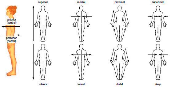
Directional terms describe the positions of one structure or part of the body relative to another part or structure of body. directional terms can be describe as shown figure shown below.

Figure 1.3 Body directions.

Term
Definition
Anterior or ventral
Nearer to front of the body or toward the front
Posterior or dorsal
After, behind or toward the back
Inferior or caudal
Below or at a lower level
Proximal
Closer to the origin
Distal
Away from the center of body
Medial
Toward the mid-line of body
Ipsilateral
On the same side of body
Deep
Away from surface of body
Superficial
 Toward or at the body surface
Intermediate
Between two structure

Table 1.1 Directional terms and explanation